CIMR Light Microscopy Facility
The primary aim of the CIMR microscopy facility is to enable high-quality research by providing the very latest and best instrumentation and imaging software to researchers at the institute. We also aim to ensure that the facility is used to maximum effect by providing comprehensive training and on-going support on all instruments and technologies available. The variety of instruments and image analysis options in the facility ensure that we are able to meet most imaging requirements in-house.
The facility is used by the majority of researchers and research groups at the CIMR and provides invaluable tools for investigating cellular processes in both fixed and living samples in multiple dimensions. This is achieved by means of conventional imaging, HCS, confocal microscopy, time-lapse, real-time widefield & TIRF, FRAP, FRET and FCS.
We are constantly assessing the requirements of the institute and developing the facility accordingly; most recently this has resulted in the expansion of our live imaging capacity with the addition of a Zeiss Elyra7 Lattice-SIM / SMLM instrument for rapid super-resolution imaging of living samples.
Our resources include:
- Widefield epi-flourescence and colour brightfield microscopes in upright and inverted configurations
- Three fully incubated laser scanning confocal microscopes variously configured with:
- Airyscan (enhanced resolution confocal imaging)
- FCS (Fluorescence Correlation Spectroscopy)
- FLIM (Fluorescence Lifetime Imaging)
- A fully incubated, super-resolution Lattice SIM (structured illumination microscopy) / SMLM (single molecule localisation microscopy) for rapid imaging of living samples
- Super-resolution microscope offering both super-resolution stripe structured illumination (SR-SIM) and localisation microscopy (e.g. PALM and STORM) for fixed samples
- High Content Screening and Analysis instrument (Thermo CellInsight CX7 with plate-handling robot)
- Image analysis workstations with leading software packages (e.g. Imaris, ZEN, FiJi, Volocity)
- Histology processing, cutting and staining equipment
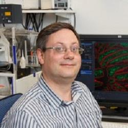
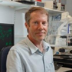
The facility is managed by two experienced microscopists, Matthew Gratian and Mark Bowen, who provide all training and support on the above instruments. Researchers external to CIMR and the University of Cambridge should contact Matthew if interested in accessing the Facility.