
Myosin motor proteins in health and disease
General audience summary:
In living cells dynamic processes such as muscle contraction and intracellular transport are powered by myosin motor proteins, which are nanomolecular machines that move cargo along actin tracks rather like a train running along a railway network to specific destinations. In our lab we follow the activity of myosin motors in living cells using high resolution microscopy but also use a wide variety of biochemical and biophysical techniques on isolated motors in the test tube to study their characteristic behaviour.
Dysfunction of motor activity is linked to many human diseases including deafness, cancer and neurodegeneration. Therefore, a deeper understanding how motors are switched on/off, fine-tuned and which cargo is transported, will guide our efforts to modulate myosin activity as a therapeutic strategy.
Strategic CIMR theme: Membrane trafficking, Organelle Biology, Intracellular Infections
Funding: Medical Research Council, Biotechnology and Biological Sciences Research Council and Wellcome Trust
Research Group Members: Sue Arden, Janeska de Jonge, Alex Holmes, Genjing Zhao
Research
Myosin motor proteins in health and disease
Intracellular transport is driven by motor proteins that use energy derived from ATP hydrolysis to organize cellular compartments and control intracellular transport along cytoskeletal tracks; defects in these fundamental transport processes are linked to a wide range of disorders including deafness, cardiomyopathy, neurodegeneration and cancer. Research in our lab is focused on the cellular function of myosin motors that generate force and move cargo along actin tracks. We are using a variety of cellular, molecular and biochemical approaches to determine how a motor recognizes and selects its cargo and how motor activity and cargo attachment are coordinated. Much of our research has focused on myosins of class I and also class VI (MYO6), a unique highly specialized class of myosin motors that move in reverse direction along actin filaments. Using in situ proximity labelling and functional proteomics we have mapped the interactome of myosins of class I and VI and identified MYO6-associated complexes that highlight the role of this myosin in regulating actin cytoskeleton dynamics. Indeed MYO6 initiates the assembly of F-actin cages around damaged mitochondria, which serves as a quality control mechanism to isolate dysfunctional organelles from the healthy network. Our overall aim is to determine the role(s) of these myosins and their cargo adaptors in cell signalling, cargo transport and cellular homeostasis and why dysfunction is linked to neurodegenerative disorders such as Alzheimer’s, Parkinson’s and motor neuron disease.
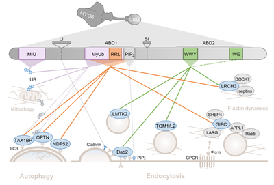
Figure 1: Cartoon highlighting binding sites in the MYO6 cargo-binding tail domain. Schematic of the cellular processes in which MYO6 is implicated through direct (blue) and indirect (grey) binding partners. (For further details see De Jonge et al., FEBS Lett. 2019)
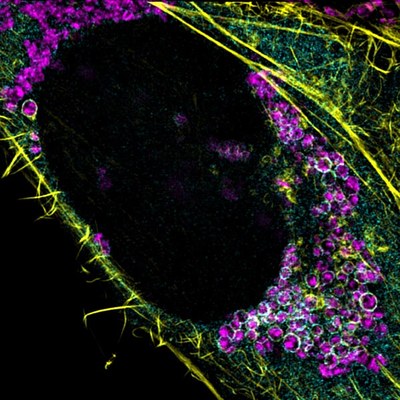
Figure 2: Mitochondrial homeostasis is maintained by removing dysfunctional, ubiquitinated mitochondria from the network via mitophagy. On the mitochondrial surface, myosin VI (cyan) initiates the assembly of F-actin cages (yellow), which serve as a quality control mechanism to isolate dysfunctional mitochondria (magenta) and thereby prevent their refusion with neighbouring populations.
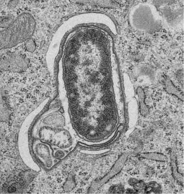
Figure 3: Xenophagy in mouse embryonic fibroblast 1 hour after infection with Salmonella. The transmission electron microscope image highlights a single bacterium surrounded by the Salmonella-containing vacuole membrane and four individual phagophores. The outer membrane of the phagophores is in close proximity to the rough endoplasmic reticulum. (Kishi-Itakura et al. Traffic 2020).
Publications
O'Loughlin T, Kruppa AJ, Ribeiro ALR, Edgar JR, Ghannam A, Smith AM, Buss F. OPTN recruitment to a Golgi-proximal compartment regulates immune signalling and cytokine secretion. J Cell Sci. 2020 Jun 15;133(12):jcs239822. doi: 10.1242/jcs.239822. PMID: 32376785; PMCID: PMC7328155.
Zakrzewski P, Lenartowska M, Buss F. Diverse functions of myosin VI in spermiogenesis. Histochem Cell Biol. 2021 Jan 2. doi: 10.1007/s00418-020-01954-x. Epub ahead of print. PMID: 33386429.
Kishi-ItakuraC, Nicholas T. Ktistakis NT, Buss F. Ultrastructural insights into pathogen clearance by autophagy. Traffic. doi: 10.1111/tra.12723 (2020).
De Jonge JJ, Batters C, O’Loughlin T, Arden SD, Buss F. The MYO6 interactome: selective motor-cargo complexes fro diverse cellular processes. FEBS Lett. 593(13): 1494-1507 (2019).
O’Loughlin, T, Masters, TA and Buss, F. The MYO6 interactome reveals adaptor complexes coordinating early endosome and cytoskeletal dynamics. EMBO Reports 19(4) doi: 10.15252/embr.201744884 (2018).
Kruppa AJ, Kishi-Itakura C, Masters TA, Rorbach JE, Grice GL, Kendrick-Jones J, Nathan JA, Minczuk M and Buss F. Myosin VI-dependent actin cages encapsulate Parkin-positive damaged mitochondria. Developmental Cell 44:489-499 (2018).
Masters TA, Tumbarello DA, Chibalina MV, Buss F. MYO6 Regulates Spatial Organization of Signaling Endosomes Driving AKT Activation and Actin Dynamics. Cell Rep. 19, 2088-2101 (2017).
Brooks AB, Humphreys D, Singh V, Davidson AC, Arden SD, Buss F, Koronakis V. MYO6 is targeted by Salmonella virulence effectors to trigger PI3-kinase signaling and pathogen invasion into host cells. Proc. Natl Acad. Sci. USA 114, 3915-3920 (2017).
Masters TA, Buss F. Filopodia formation and endosome clustering induced by mutant plus-end-directed myosin VI. Proc. Natl Acad. Sci. USA 114, 1595-1600 (2017).
Tumbarello DA, Manna P, Allen M, Bycroft M, Arden SD, Kendrick-Jones J & Buss F. The autophagy receptor TAX1BP1 and the molecular motor myosin VI are required for clearance of Salmonella typhimurium by autophagy. PLoS Pathogens 11, e1005174. doi: 10.1371/journal.ppat.1005174 (2015).
Brandstaetter, H, Kishi-Itakura, C, Manstein D, Tumbarello D. and Buss F. Loss of functional myosin 1c, a motor protein involved in lipid raft trafficking, disrupts autophagosome-lysosome fusion. Autophagy 10, 2310-23 (2014).
Tumbarello, D. A., Waxse, B. J., Arden, S. D., Bright, N. A., Kendrick-Jones, J. and Buss, F. Autophagy-receptors link myosin VI to autophagosomes to mediate Tom1-dependent autophagosome maturation and fusion with the lysosome. Nature Cell Biol. 10, 1024–1035 (2012).