
Our facility supports a range of structural and computational research laboratories using X-ray crystallography, cryo electron microscopy, molecular modelling and new method development. Our structural biology research is providing new insights into the molecular mechanisms underlying a range of human diseases. Our users combine structural work with complementary methods in cell biology, biochemistry and biophysics to develop a comprehensive understanding of fundamental biological processes.
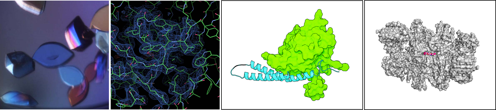
Protein Crystallography
The facility has high-throughput robotics for protein crystallisation including:
Mosquito LCP Crystallisation Robotics for nanoscale drop setting
Formulatrix Rock Imager for dynamic monitoring of protein crystallisation:
We have regular access to synchrotron light sources including Diamond Light Source for remote access data collection and data processing.
Cryo-electron microscopy
Within the CIMR we have access to infrastructure for the preparation of cryo-EM samples including:
Vitrobot for plunge freezing cryo-EM samples
For data collection we have access to the electron microscope facility in the Department of Biochemistry.
Software Development
Within CIMR we have a research team dedicated to the development of new computational methods for the improvement of structure determinaton and refinement.
Phaser is a program for phasing macromolecular crystal structures using maximum likelihood methods and is part of the Phenix and CCP4 software suites for structural biology.
ISOLDE is an interactive environment designed to ease the task of building high-fidelity atomic models in challenging low-resolution crystallographic and cryo-EM datasets by making use of GPU-accelerated interactive molecular dynamics combined with real-time geometric validation.
Selected recent publications
Terwilliger TC, Ludtke SJ, Read RJ, Adams PD and Afonine PV. Improvement of cryo-EM maps by density modification. Nature Methods 17, 923-927 (2020).
Hay IM, Fearnley GW, Rios P, Köhn M, Sharpe HJ, Deane JE. The receptor PTPRU is a redox sensitive pseudophosphatase. Nature Communications 11:3219 (2020).
Butt BG, Owen DJ, Jeffries CM, Ivanova L, Hill CH, Houghton JW, Ahmed MF, Antrobus R, Svergun DI, Welch JJ, Crump CM, Graham SC Insights into herpesvirus assembly from the structure of the pUL7:pUL51 complex. eLife 9: e53789 (2020).
Rougé L, Chiang N, Steffek M, Kugel C, Croll TI, Tam C, Estevez A, Arthur CP, Koth CM, Ciferri C, Kraft E, Payandeh J, Nakamura G, Koerber JT, Rohou A. Structure of CD20 in complex with the therapeutic monoclonal antibody rituximab. Science 367:1224-1230 (2020).
Wrobel AG, Kadlecova Z, Kamenicky J, Yang JC, Herrmann T, Kelly BT, McCoy AJ, Evans PR, Martin S, Müller S, Salomon S, Sroubek F, Neuhaus D, Höning S, Owen DJ. Temporal Ordering in Endocytic Clathrin-Coated Vesicle Formation via AP2 Phosphorylation. Developmental Cell 50:494-508.e11 (2019).
Perera LA, Rato C, Yan Y, Neidhardt L, McLaughlin SH, Read RJ, Preissler S, Ron D. An oligomeric state-dependent switch in the ER enzyme FICD regulates AMPylation and deAMPylation of BiP. EMBO J.:e102177 (2019).
Gao C, Pallett MA, Croll TI, Smith GL, Graham SC. Molecular basis of cullin-3 ubiquitin ligase subversion by vaccinia virus protein A55. Journal of Biological Chemistry, 294: 6416–6429 (2019).
Yan Y, Rato C, Rohland L, Preissler S and Ron D. MANF antagonizes nucleotide exchange by the endoplasmic reticulum chaperone BiP. Nature Communications 10:541 (2019)
Zyryanova AF, Weis F, Faille A, Alard AA, Crespillo-Casado A, Sekine Y, Harding HP, Allen F, Parts L, Fromont C, Fischer PM, Warren AJ, Ron D. Binding of ISRIB reveals a regulatory site in the nucleotide exchange factor eIF2B. Science 359:1533-1536 (2018).
Hill CH, Cook GM, Spratley SJ, Fawke S, Graham SC, Deane JE. The mechanism of glycosphingolipid degradation revealed by a GALC-SapA complex structure. Nature Communications 9:151 (2018).
Croll TI. ISOLDE: a physically realistic environment for model building into low-resolution electron-density maps. Acta Crystallogr D Struct Biol. 74:519-530 (2018).