Control of secretion at the immunological synapse
General audience summary:
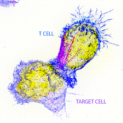
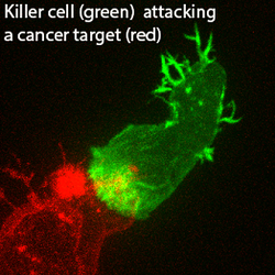
Within every teaspoon of our blood are 5 million potential killer cells. These are remarkable little cells that circulate around our bodies recognizing and destroying virally infected and cancer cells with extraordinary specificity and precision. We have discovered new genes that suggest that many different components within killer cells play a part in delivering the lethal hit. We wish to work out how each part of the killer cell contributes. We study killer cells in which the new genes are missing to see where things go wrong. We are able to film live killer cells and pin-point what is different in each case. This is particularly important right now as new cancer therapies in medicine aimed at helping killer cells are proving very effective. By understanding exactly how killer cells work, it will be possible to devise better therapies.
Strategic CIMR themes: Intracellular Infections, Organelle Biology, Rare Genetic Diseases
Research Group Members: Yukako Asano, Claire Ma, Martin Limback-Stokin, Gurpreet Dhaliwal, Megan Glover, Gordon Frazer, Yuxuan Lin (Kaite)
Research
Control of secretion at the immunological synapse
Cytotoxic T lymphocytes (CTLs) play a critical role in the immune system, recognizing and destroying virally infected cells and cancer targets with remarkable specificity. These cells are extraordinarily efficient serial killers that rapidly deliver their lethal hit using precisely polarized secretion of cytolytic proteins from modified lysosomes to destroy their targets. CTLs provide a fascinating system in which to understand the cell biology of secretion in a specialized cell type. This is particularly important right now, with new immunotherapies focused on harnessing the cytotoxic potential of these cells to combat cancer.
The ability to grow primary CTLs in culture from <5ml of blood has been very powerful, allowing us early on to use genetics to understand function by generating CTLs from patients with genetic mutations. Using biochemical, molecular and high-resolution imaging approaches we have made many fundamental discoveries both in the field of cytotoxicity as well as cell biology, identifying proteins required to make a lysosome a secretory organelle and describing a novel role for the centrosome in CTL secretion revealing unexpected parallels with ciliogenesis.

Our high resolution, temporal imaging has pinpointed the timing and location of key events leading to secretion with increasing detail emerging with each new probe examined. With the ability to express tagged or modified proteins in CTLs together with CRISPR technology to generate mutations by design, it is now possible to probe CTL function in greater detail than ever before.
Link to live imaging of T cells killing target cells (on YouTube)
Publications
Barton PR, Davenport AJ, Hukelmann J, Cantrell DA, Stinchcombe JC and Griffiths GM. Super-killer CTLs are generated by single gene deletion of Bach2. European Journal of Immunology, Epub ahead of print. doi: 10.1002/eji.202249797 (2022).
Lisci M, Griffiths GM. Arming a killer: mitochondrial regulation of CD8+ T cell cytotoxicity. Trends in Cellular Biology, Epub ahead of print. doi: 10.1016/j.tcb.2022.05.007 (2022).
Richard AC, Frazer G, Ma CY, Griffiths GM. Staggered starts in the race to T cell activation. Trends in Immunology, 42(11):994-1008. doi: 10.1016/j.it.2021.09.004 (2021)
Frazer G, Gawden-Bone CM, Dieckmann NMG, Asano Y and Griffiths GM. Signal strength controls the rate of polarization within CTLs during killing. Journal of Cell Biology, 220 (10): e202104093 (2021).
Lisci M, Barton PR, Randzavola LR, Ma CY, Marchingo JM, Cantrell DA, Paupe V, Prudent J, Stinchcombe JC and Griffiths GM. Mitochondrial translation is required for sustained killing by cytotoxic T cells. Science, 15;374(6565):eabe9977. doi: 10.1126/science.abe9977 (2021).
Ma, C, Marioni, J, Griffiths, GM and Richard, A. Stimulation strength controls the rate of initiation but not the molecular organization of TCR-induced signalling. eLife, 9:e53948. doi: 10.7554/eLife.53948 (2020).
Randzavola LO, Strege K, Juzans M, Asano Y, Stinchcombe JC, Gawden-Bone CM, Seaman MNJ, Kuijpers T. and Griffiths GM. Loss of ARPC1B impairs cytotoxic T lymphocyte maintenance and cytolytic activity. Journal of Clinical Investigation, 129(12), pp.5600-5614 (2019).
Ritter AT, Kapnick SM, Murugesan S, Schwartzberg PL, Griffiths GM, Lippincott-Schwartz J. Cortical actin recovery at the immunological synapse leads to termination of lytic granule secretion in cytotoxic T lymphocytes. Proc. Natl Acad. Sci. USA 114(32):E6585-E6594. doi: 10.1073/pnas.1710751114 (2017).
Stinchcombe JC, Randzavola L, Angus KL, Mantell JM, Verkade P & Griffiths GM. Mother Centriole Distal Appendages Mediate Centrosome Docking at the Immunological Synapse and Reveal Mechanistic Parallels with Ciliogenesis. Current Biol. 25, 3239–3244 (2015).
Ritter AT, Asano Y, Stinchcombe JC, Dieckmann NM, Chen BC, Gawden-Bone C, van Engelenburg S, Legant W, Gao L, Davidson MW, Betzig E, Lippincott-Schwartz J, Griffiths GM. Actin depletion initiates events leading to granule secretion at the immunological synapse. Immunity 42, 864-876. doi: 10.1016/j.immuni.2015.04.013 (2015).
Jenkins, M. R. et al. Distinct structural and catalytic roles for Zap70 in formation of the immunological synapse in CTL. eLife 3:e01310 (2014).
de la Roche, M. et al. Hedgehog signaling controls T-cell killing at the immunological synapse. Science 342, 1247–1250 (2013).